MACS Matchmaker
The AminoLink DDI strategy conjugates a protein's primary amines (lysines or N-terminus) to a TCO-functionalized oligonucleotide off-chip with the Oligonucleotide Conjugation Kit, AminoLink, then hybridizes the resulting protein–oligo conjugate to any Oligo|PEG chip via DNA-Directed Immobilization (DDI). The duplex is the weakest link, so the chip is regenerable between different ligands by an alkaline strip. Validated on antibodies, Fabs, nanobodies, Fc receptors, and other recombinant proteins.
Before you start
- Read Safety & Compliance and the safety data sheet for every reagent below. The SDS, not this page, governs how a reagent is handled, stored and disposed of.
- Use the personal protective equipment and the waste route your laboratory prescribes for these reagent classes.
- Check this protocol against the materials actually in hand: confirm the intended Seq ID, the product revision, the lot-specific certificate and the instructions supplied with it. Where those differ from this page, the documentation supplied with the material governs.
From the catalogue
Order the kit and the matching adapter from the lino Biotech webshop:
| Item | Contents | Webshop |
|---|---|---|
| Oligonucleotide Conjugation Kit, AminoLink | Protein Preparation Buffer, Me-Tz bifunctional linker with its reconstitution medium, desalting column, oligonucleotide capturing slurry with spin column, protein concentrator and collection tubes; one conjugation of 100 µg of protein (15–300 kDa) in under 4 h | View in shop |
| Oligo Adapter for Amine Kit | Lyophilised, 10 nmol; TCO-modified ssDNA, one Seq ID (01–64) per vial; reconstitute at 100 µM; store at −20 °C | View in shop |
| Desalting Column, Zeba 7K | Spare for the excess-linker removal step; one is included in the kit | View in shop |
| Spin Column, Amicon 10K | Spare protein concentrator for the optional concentration or buffer exchange (not the slurry spin column); one is included in the kit | View in shop |
Strategy: two-step off-chip coupling
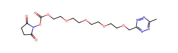
Conjugation proceeds in two reactions in solution, each followed by a clean-up on a kit column:
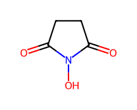
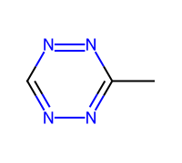
- Me-Tz linker activation. The NHS ester of the bifunctional linker reacts with a primary amine on the protein, installing a methyltetrazine (Me-Tz) handle. The linker volume sets the average number of handles per protein, from one to five.
- Removal of excess linker. The desalting column retains free linker, which would otherwise consume TCO-oligo in the next step.
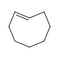
- TCO-oligo click. The Me-Tz handle reacts with the TCO-functionalized oligo via inverse-electron-demand Diels–Alder (IEDDA). The oligo volume follows the same protein-to-oligo ratio.
- Removal of free oligo. The oligonucleotide capturing slurry retains free oligo while the conjugate passes through.
Protocol
Step 1 — Protein requirements and optional buffer exchange
Start from 100 µg of protein (15–300 kDa) at 1–5 mg/mL, free of carrier proteins such as BSA. The NHS ester reacts with any primary amine, so the formulation must meet the requirements below; exchange the buffer first if it does not, or if the protein is below 1 mg/mL. Reconstitute lyophilised protein at 1 mg/mL in Protein Preparation Buffer.
| Protein formulation | Requirement |
|---|---|
| pH | 7–8 |
| Amine-free buffer (e.g. PBS) | Allowed |
| Non-buffering salts, chelating agents (EDTA), sugars | Allowed |
| Glycerol | ≤ 40 % (v/v) |
| Sodium azide | ≤ 0.05 % (w/v) |
| Tris, glycine, histidine, other primary amines | Not allowed |
To exchange the buffer, wash the protein concentrator with 500 µL Protein Preparation Buffer at 14,000 × g for 10 min, bring the protein to 450 µL with Protein Preparation Buffer, spin at 14,000 × g for 10 min and discard the flow-through, then refill with 450 µL buffer and spin again. Repeat the refill as many times as the amine-containing component needs: the number of exchanges is the decimal logarithm of its concentration in µM, rounded up (20 mM Tris needs five). Invert the concentrator into a fresh collection tube and spin at 1,000 × g for 2 min; about 50 µL at about 2 mg/mL is recovered.
Step 2 — Reconstitute the linker
Add 10 µL Linker Reconstitution Medium to the bifunctional linker and pipette vigorously until the red pellet has dissolved. Add 2.0 mL Protein Preparation Buffer, vortex for at least 5 s and spin down. Use the solution immediately.
Step 3 — Activate the protein
Add the linker to the solution containing 100 µg of protein. The volume follows from the protein's molecular weight and the number of oligos wanted per protein:
linker volume (µL) = oligos per protein ÷ molecular weight (kDa) × 1000
| Protein (kDa) | 1 : 1 | 1 : 2 | 1 : 5 |
|---|---|---|---|
| 15 | 70 µL | — | — |
| 50 | 20 µL | 40 µL | 100 µL |
| 100 | 10 µL | 20 µL | 50 µL |
| 150 | 7 µL | 14 µL | 35 µL |
Incubate in a thermomixer at 25 °C and 800 rpm for 1 h. The ratio is a statistical average: most proteins carry the chosen number of handles, individual molecules carry more or fewer.
Step 4 — Remove excess linker
Remove the bottom closure of the desalting column, loosen its cap, place it in a 2 mL collection tube and spin at 1,500 × g for 1 min to remove the storage solution. Mark the side where the resin slants upward and keep that mark facing outward in every following spin. Equilibrate with 300 µL Protein Preparation Buffer at 1,500 × g for 1 min, three times. Bring the activated protein to 130 µL with Protein Preparation Buffer, apply it slowly to the centre of the resin and collect it at 1,500 × g for 2 min.
Step 5 — Conjugate to the TCO-oligo
Reconstitute the adapter at 100 µM in nuclease-free water, PBS or Protein Preparation Buffer. Add it to the purified activated protein; the volume follows the same ratio as the linker:
oligo volume (µL) = oligos per protein ÷ molecular weight (kDa) × 1500
| Protein (kDa) | 1 : 1 | 1 : 2 | 1 : 5 |
|---|---|---|---|
| 15 | 100 µL | — | — |
| 50 | 30 µL | 60 µL | 150 µL |
| 100 | 15 µL | 30 µL | 75 µL |
| 150 | 10 µL | 20 µL | 50 µL |
Incubate in a thermomixer at 25 °C and 800 rpm for 1 h.
Step 6 — Remove free oligo
Vortex the oligonucleotide capturing slurry briefly and add 200 µL to the spin column. In a cap-free collection tube, spin at 700 × g for 2 min, then equilibrate with 200 µL Protein Preparation Buffer at 700 × g for 2 min, three times. Bring the conjugation mixture to 200 µL with Protein Preparation Buffer, close the bottom of the spin column, place it in a fresh 2 mL collection tube and apply the sample slowly to the centre of the slurry. Incubate in a thermomixer at 25 °C and 800 rpm for 30 min. Remove the bottom closure and spin at 700 × g for 2 min; add 200 µL Protein Preparation Buffer and spin again to recover the rest. The collection tube now holds the purified conjugate.
The final concentration depends on the protein and the ratio used; measure it with an appropriate assay where it matters. Aliquot and store at +4 °C for up to 3 months.
Step 7 — Hybridisation to chip and measurement
| Parameter | Value |
|---|---|
| Ligand concentration | 200 nM in PBST |
| Flow rate | 10 µL/min |
| Contact time | 5 min |
| Temperature | Ambient (the reader holds no fixed set point) |
| Chip type | Oligo|PEG (any plex count) |
| SCK association flow rate | 30 µL/min (6-ch) / 60 µL/min (3–4-ch) / 100 µL/min (1-ch) |
| SCK dissociation flow rate | 200 µL/min |
| Regeneration (duplex strip) | 3 M GuHCl + 125 mM NaOH, 400 µL/min, 0.5 min |
Reference protocol parameters
| Parameter | Reference value |
|---|---|
| Protein input | 100 µg at 1–5 mg/mL, 15–300 kDa, amine-free buffer pH 7–8 |
| Oligos per protein | 1 to 5, set by the linker and oligo volumes |
| Activation | 25 °C, 800 rpm, 1 h |
| Excess-linker removal | Desalting column, sample in 130 µL, 1,500 × g, 2 min |
| Conjugation | TCO-oligo at 100 µM, 25 °C, 800 rpm, 1 h |
| Free-oligo removal | Capturing slurry, sample in 200 µL, 30 min at 800 rpm, 700 × g |
| Storage of the conjugate | +4 °C, up to 3 months |
| Hybridisation concentration | 200 nM |
| Hybridisation flow rate | 10 µL/min, 5 min |
| SCK association flow rate | 30 µL/min (6-ch) / 60 µL/min (3–4-ch) / 100 µL/min (1-ch) |
| SCK dissociation flow rate | 200 µL/min |
| Regeneration | 3 M GuHCl + 125 mM NaOH, 400 µL/min, 0.5 min |
QC and acceptance
Before the conjugate goes on a chip, establish that the ligand is actually carrying oligo. The conjugation is the step that fails silently: an unconjugated ligand behaves exactly like a ligand that never bound its target.
- Off-chip. A mobility shift against the unconjugated ligand on SDS-PAGE, or the corresponding shift by SEC, shows that oligo is attached. An A260/A280 ratio raised above the ligand's own value is consistent with the same thing and is quicker, but it does not distinguish attached oligo from free oligo carried through the purification.
- On-chip. The conjugate injection gives an immobilization response that survives the following buffer wash, and a mologram carrying a non-complementary strand does not. That pair separates hybridization from adsorption.
There is no validated degree-of-conjugation threshold for this protocol. Record the value you measure and keep it comparable across batches rather than judging it against a number.
Practical notes
See also: immobilization method comparison table.